

what structures pass though the supra orbital foramen, infra orbital foramen and mental foramen?
Anterior view
Supra-orbital foramen Supra-orbital nerve and vessels (branch of frontal nerve which is branch of opthalamic nerve trigeminal)
Infra-orbital foramen Infra-orbital nerve and vessels (maxillary nerve, but in the canal called that way)
Mental foramen Mental nerve and vessels (branch of mandibular division of trigeminal)
ALL OF THEM BRANCHES OF TRIGEMINAL
on what sides can facial paralzsis occur?
what does a lower motor lesion to in the facial musculature?
what can damage to facial nerve cause?
what does a lower motor lesion to in the facial musculature?
what can damage to facial nerve cause?
the internal acoustic meatus (by a tumour),
in the middle ear (by infection or surgery),
facial nerve canal (Bell’s palsy),
in the parotid gland (tumour)
through laceration or injury to
the face.
drooping of the lower eyelid and the angle of the mouth.
paralysis of the orbicularis oculi muscle leading to incomplete eyelid closure during both voluntary and reflex movements.
Eyelid closure is the means by which the cornea is lubricated; functional deficits in the ability to close the
eyelid can therefore lead to corneal damage and permanent vision impairment.
what movements does the buccinator control?
what does it serve?
what causes its paralysis?
what does it serve?
what causes its paralysis?
• It controls the cheeks in blowing and sucking (an important muscle in the newborn).
• It serves to empty the gutter between the teeth and cheek during mastication.
• Paralysis of buccinator causes food to collect between the cheeks and teeth and an inability
to whistle.
• Note that the duct of the parotid gland pierces buccinator.
form where does the facial come?
through what kan it easily be recognised?
it is together with the transverse facial the major blood supplz for what?
through what kan it easily be recognised?
it is together with the transverse facial the major blood supplz for what?
external carotid artery, behind the angle of the mandible, looping behind the mandible first before crossing the lower border of the mandible to pass upwards and medially towards the medial angle of the eye.
• The facial artery, along with the transverse facial artery, is a major artery supplying the
superficial face
• The facial artery is tortuous, especially near the angle of the mouth (to allow for stretch
when the mouth is opened)
• The facial artery, along with the transverse facial artery, is a major artery supplying the
superficial face
• The facial artery is tortuous, especially near the angle of the mouth (to allow for stretch
when the mouth is opened)
what are the 5 branches of the facial arterz and what do they supplz?
Branches of the facial artery (see Figure 2.1) include:
• Inferior labial artery (to the lower lip)
• Superior labial artery (to the upper lip)
• Muscular branches (to the muscles of facial expression)
• Lateral nasal artery (to the ala and dorsum of nose)
• Angular artery
• Inferior labial artery (to the lower lip)
• Superior labial artery (to the upper lip)
• Muscular branches (to the muscles of facial expression)
• Lateral nasal artery (to the ala and dorsum of nose)
• Angular artery
name the 3 major branches of the maxillarzy arterz and what they supply/.
Maxillary Artery
Facial branches of the maxillary artery include:
• Infraorbital artery (lower eyelid, lateral aspect of nose and upper cheek)
• Buccal artery (cheek)
• Mental artery (terminal branch of inferior alveolar (of maxillary) which supplies muscles
and skin of chin)
Facial branches of the maxillary artery include:
• Infraorbital artery (lower eyelid, lateral aspect of nose and upper cheek)
• Buccal artery (cheek)
• Mental artery (terminal branch of inferior alveolar (of maxillary) which supplies muscles
and skin of chin)
where does the superficial tempral artery arise in what structure?
what are its bracnhes?
what does it supplz?
what does its main branch supplz? 4
what are its bracnhes?
what does it supplz?
what does its main branch supplz? 4
• small terminal branch of external carotid that arises within parotid gland
• gives off several branches (parotid, frontal, parietal, middle temporal and transverse facial arteries) to
- supply the lateral aspect of the scalp• the transverse facial artery is one of the main arteries of the superficial face. It crosses masseter above the parotid duct and supplies the parotid gland, parotid duct, masseter
and skin.
what are the chief facial veins? 3
what is the problem with them?
what is the problem with them?
facial vein retromandibular and superficial temporal vein.
- valveless, and pressure or blockage can therefore cause blood to flow into the cavernous sinus.
- facial vein sommunicates with the cranial cavitz via ophthalmic veins and the pterygoid plexus
-infection from a danger area (e.g. pimple on nose) can spread to the cavernous sinus with grave consequences (cavernous sinus thrombosis).
list the blood supplz of the scalp. there are to major arteris suplzing the sclap. list them and the major branches and thwat they supplz. one has 3 and one has 2 major branches.
1. External carotid* (gives three branches to scalp)
i. Occipital artery supplies the posterior scalp
ii. Posterior auricular artery supplies scalp posterior to the ear
iii. Superficial temporal artery supplies the lateral scalp
2. Ophthalmic artery (a branch of the internal carotid* gives two branches to the scalp)
i. Supratrochlear artery supplies the anteromedial scalp
ii. Supraorbital artery supplies the anterolateral scalp
list the MAIN trigeminal branches supplzing stuff to the face.
a. Ophthalmic division (nerve) – gives two branches to the scalp:
- Supratrochlear (anteromedial portion of scalp)
- Supraorbital (anterolateral portion of scalp)
b. Maxillary division (nerve) – gives one branch to scalp
- Zygomaticotemporal nerve (supplies temporal region)
c. Mandibular division (nerve) – gives one branch to scalp
- Auriculotemporal nerve (supplies temporal region)
what structures do we find in the connective tissue?
what woud happen if you wuld have a wound in it and why/
what woud happen if you wuld have a wound in it and why/
• Contains arteries, veins and nerves
(highly vascular)
• Scalp wounds bleed profusely because severed blood vessels cannot contract – their walls are held open by dense connective
tissue.
.
(highly vascular)
• Scalp wounds bleed profusely because severed blood vessels cannot contract – their walls are held open by dense connective
tissue.
.
the epicranial aponeurosis attaches from ewhere to where?
what would happen if a wound would penetrate this layer?
what would happen if a wound would penetrate this layer?
external occipital protuberance and nuchal line to the occipital and frontal bellies of occipitofrontalis
• A wound that penetrates this layer gapes, since the aponeurotic layer is pulled apart by the contraction of the bellies of occipitofrontalis.
• A wound that penetrates this layer gapes, since the aponeurotic layer is pulled apart by the contraction of the bellies of occipitofrontalis.
what does the loose connective tissue contain and what is special about it?
what does this specialness mean clinically?
what does this specialness mean clinically?
• Contains a few small arteries, and emissary veins. These veins are valveless and connect superficial veins of scalp with the diploic veins of skull bones and with the intercranial venous sinuses.
:
:
what is the difficulitz with the loose connective layer?
- Infection can easily spread in it and may enter the cranial cavity via emissary veins
- Fluid and pus may accumulate here
- It is the site of extracranial haematoma (bleeding in the subaponeurotic space)
- Looseness of the connective tissue causes blood in the subaponeurotic space to track
laterally to the temporal lines, posteriorly to the superior nuchal line and anteriorly to
the upper (and to a lesser degree the lower) eyelids to give a black eye.

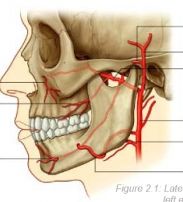
froom upper left to right anti clock wise
supratrochlear artery and vein
supraorbital artery and vein
angular vein and artery
superior labial vein and artery
infeiror labial vein and artery
faical artery
facial vein
ezternal carotid
internal jugular
external jugular
occipital artery
occipital vein
posterior aruicular artery
posterior auricular vein
superficial temporal artery and vein
transverse facial artery and vein
zygomaticofcial artery and vein (the one down)
zygomaticotemporal artery and vein
the obicularis oculi is seperated into three parts. name them and their action.
orbital part: causes the eyelid to close tightlz
palpebral part: cuases to close the eyelid gently
lacrimal part: attaches to lacrimal saz and contraction pulls the lacrimal sac open and kkeeps the lacirmal puncta in contact with the eyeball.
palpebral part: cuases to close the eyelid gently
lacrimal part: attaches to lacrimal saz and contraction pulls the lacrimal sac open and kkeeps the lacirmal puncta in contact with the eyeball.
obicularis oculi has three parts. what are their actions?
• Orbital part – concentric fibres which sweep around the orbit; contraction of these fibres causes you to close your eyelids tightly.
• Palpebral part – fibres which pass across the eyelid; contraction of these fibres causes you to close your eyelids gently (blinking / sleeping).
• Lacrimal part – a few fibres attach to the lacrimal sac; contraction pulls the lacrimal sac open and keeps the lacrimal puncta in contact with the eyeball.
Occipitofrontalis and levator palpebrae superioris antagonise the action of orbicularis ocul
obicularis oris is a shincter of the mouth. what are its actions? what are its anagonists 10?
• This muscle is a sphincter of the mouth, it acts to narrow the mouth and close the lips
(
Antagonists of orbicularis oris are dilators of the mouth in various directions – these are:
• Levator labii superioris alaeque nasi
• Levator labii superioris
• Zygomaticus minor
• Zygomaticus major
• Levator anguli oris
• Risorius
• Depressor anguli oris
• Depressor labi inferioris
• Mentalis
Buccinator

what are the branches of the facial artery? 5 the facial artery supplis with the transverse facial most of the stuff int eh face.
Branches of the facial artery (see Figure 2.1) include:
• Inferior labial artery (to the lower lip)
• Superior labial artery (to the upper lip)
• Muscular branches (to the muscles of facial expression)
• Lateral nasal artery (to the ala and dorsum of nose)
• Angular artery
• Inferior labial artery (to the lower lip)
• Superior labial artery (to the upper lip)
• Muscular branches (to the muscles of facial expression)
• Lateral nasal artery (to the ala and dorsum of nose)
• Angular artery

what are the branches of the maxillary artery?
Facial branches of the maxillary artery include:
• Infraorbital artery (lower eyelid, lateral aspect of nose and upper cheek)
• Buccal artery (cheek)
• Mental artery (terminal branch of inferior alveolar (of maxillary) which supplies muscles
and skin of chin)
• Infraorbital artery (lower eyelid, lateral aspect of nose and upper cheek)
• Buccal artery (cheek)
• Mental artery (terminal branch of inferior alveolar (of maxillary) which supplies muscles
and skin of chin)
Frage
1 supratrochlear artery and vein
2 supraorbital artery and v
3 angular artery and v
4 lateral nasal artery and v
5 superir labial artery and v
6 inferior labial artery v
7 facial a
8 facial v
9 external caoritd
10 jugular vein
11 external jugular vein
12 occipital a
13 occipital v
14 posterior auricular a
15 post auricular v
16 usperficial temproal a and v
17 transverse facial a and veiin
18 zygomatical facial a and v
19 zygomatical temproal a andv
superficial temporal a arises within what structure?
where can u feel its pulse?
what are its branches? and what do they supply?
what is the main artery it gives off? and what does this one supply?
where can u feel its pulse?
what are its branches? and what do they supply?
what is the main artery it gives off? and what does this one supply?
parotid gland
•pulse felt over zygomatic arch
• gives off several branches (parotid, frontal, parietal, middle temporal and transverse
facial arteries) to supply the lateral aspect of the scalp
• the transverse facial artery is one of the main arteries of the superficial face. It crosses masseter above the parotid duct and supplies the parotid gland, parotid duct, masseter
and skin.
what are the two divison of the othalamic nerve?
the one of the macillary?
the one of the mandivular division?
know them!
the one of the macillary?
the one of the mandivular division?
know them!
Trigeminal Branches
a. Ophthalmic division (nerve) – gives two branches to the scalp:
- Supratrochlear (anteromedial portion of scalp)
- Supraorbital (anterolateral portion of scalp)
b. Maxillary division (nerve) – gives one branch to scalp
- Zygomaticotemporal nerve (supplies temporal region)
c. Mandibular division (nerve) – gives one branch to scalp
- Auriculotemporal nerve (supplies temporal region)
obicularis oris is a shincter of the mouth. what are its actions? what are its anagonists 10?
• This muscle is a sphincter of the mouth, it acts to narrow the mouth and close the lips
(
Antagonists of orbicularis oris are dilators of the mouth in various directions – these are:
• Levator labii superioris alaeque nasi
• Levator labii superioris
• Zygomaticus minor
• Zygomaticus major
• Levator anguli oris
• Risorius
• Depressor anguli oris
• Depressor labi inferioris
• Mentalis
Buccinator
what are the four hallmarks of the skin on the scalp?
connective tissue 4 ?
aponeurotic layer (or called epicranial aponeurosis) 2 ?what does this layer cover?
pericranium 3?
connective tissue 4 ?
aponeurotic layer (or called epicranial aponeurosis) 2 ?what does this layer cover?
pericranium 3?
thin (exept occipital region)
manz sebacceous glands and hair follicles
good arterial and venous supply
good lympathatic drainage
thick
highlz vasularised
many cutaneous nerves
dnese
tendinous sheet
provides attachement for occipitofrontalis, superior auricular
covers calvaria between occipitalis posterior, superior auricular laterally and anteriorlly of the forntal belly of the occipitalis (frontalis)
dense layer of connective tissue
forms periosteum of calvaria
firmly attached

1 opthalamic nerve (exists trhough superior orbital frissure)
2 maxillary nerve (v2( (formaen rotundum)
3 auriculotemproal (v3) (external acoustic meatus, the surface of the tympanic membrane (eardrum), and a large area of the temple)
4 mandibular (v3, formann ovale)
5 zygomaticofacial (v2) (small area of skin over the zygomatic bone
6 mental nerve(v3) skin and the mucous membrane of the lower lip and skin of chin
7 buccal nerve (v3) (buccinator muscle supplying the cheek)
8 infraorbital nerve (V2) (INFRAORBITAL foramen, lower eyelid, cheek, side of the nose, and upper lower eyelid, cheek, side of the nose, and upper)
9 external nasal nere (1) (anterior part of the nose)
10 infratrochlear nerve (V1) (medial half of the upper eyelid, the skin in the area of the medial angle, and the side of the nose;
11 lacrimal nerve (v1) (lateral half of the upper eyelid and the skin in the area of the lateral angle)
12 supratrochlear nerve (v1) (upper eyelid, forehead, and scalp)
13 suspraorbital nerve (V1) (upper eyelid, forehead, and scalp)
14 zygomaticotemproal nerve (v2) ( anterior temple above the zygomatic arch;)


name the structures (10 not importat)
what does pass though structure 11 ?

what does pass though structure 11 ?
1. Mandibular Condyle
2. Mandibular Notch
3. Coronoid Process
4. Ramus
5. Angle
6. Oblique Line
7. Body
8. Alveolar Process
9. Mental Foramen
11. Mandibular Foramen
mandibular nerve is one of three branches of the trigeminal nerve (CN V) and the only branch with motor innervation.
The inferior alveolar nerve
inferior alveolar artery enter the foramen traveling through the body and exit at the mental foramen on the anterior mandible at which point the nerve is known as the mental nerve.


1 frontal bone
2 gabella
3 nasion (articulation of nasal bone with frontal bone)
4 piriform aperature
5 inferoir nasal concha
6 zygomatic process of maxilla
7 ramus of mandible
8 maxilla
9 angle of mandible
10 body of mandible
11 metnal protubernace
12 menal tubercle
13 mental foramen
14 mandible
15 alevolar part of mandible
16 obliqze line
17 alevolar provesses
18 anterior nasal spine
19 nasal crest
20 infra orbiat formaen
21 zygomatic bone
22 frontal process of maxilla
23 nasal bone
24 zygomatic process of frontal bone
25 supra orbital noth
26 supracillary arch

1 coronal suturre
2 shenoparietal suture
2A shpenosqamous stuture
3 sqamous suture
4 spamous part of temporal bone
5 parietal bone
6 parietomastoid suture
7 lamboid suture
8 asterion (end of parietomastoid suture)
9 occipital bone
10 occipital mastoid suture
11 mastoid part of temporal bone
12 mastoid process
13 tympanic part of temporal bone
14 styloid rocess
15 condylar process
16 angle
17 ramus of manible
18 zsgomatic process of temporal bone
19 coronoid
20 temoral process of zygomatic bone
21 body of mandible
22 mental foramen
23 aloevolar part of manible
24 maxilla
25 zygomatic bone
26 zygomaticofacial foramen
27 nasal bone
28 lacrimal bone
29 greater wing of sphenoid
30 forntal bone
31 pterion


1 hard pallate (maxilla)
2 hard palate (palantine bone)
3 greater palatine froamen
4 lesser palatine foramen
5 body of sphenoid
6 medial process of pterygoid process
7 lateral process of pterygoid process
8 scaphoid fossa
9 formaen lacerum
10 foramen ovale
11 foramen spinosum
12 carotid canal
13 stylomastoid foramen
14 basilar part of occipital bone
15 pharyngeal tubercle
16 inferoir nuchal line
17 sup nuchal line
18 external occipital protuberance
18 a extenral occipital crest
19 foramen magnum
20 occpital condyle
21 hypoglossal canal
22 mastoid notch
23 mastoid process
24 jugular foramen
25 styloid process
26 groove for auditory tube
27 mandibular fossa
28 articular tubercle
29 opening of pterygoid canal
30 greater wing
31 vomer
32 pterygoid fossa
33 pyramindal process of palatine bone
34 hamulus
35 posterior nasal aperture (choana)
36 alevolar arch
37 posterior nasal spione
38 insisive fossa


1 middle clinoid process
2 optic canal
3 foramen rotundum
4 superior orbital fissure
5 greater wing
6 openign of carotid canal
7 grrove for middle menigeal artery
8 foramen spinosum
9 foramen ovale
10 foramen lacerum
11 dorsum sellae
12 groove and hiatus of greater petrosal nerve
13groove and hiatus for lesser petrosal nerve
14 psoterior clinoid process
15 hypophyseal fossa
16 tuberculum sellae
17 chiasmatic sulcus

1 clivus (boundary of an. part of post. cranial fossa)
2 jugular tubercle
3 internal acoustic meatus
4 jugular foramen
5 hypoglossal foramen
6 foramen magnum
7 internal occipital protuberance
8 internal occipital crest
9 groove for transverse sinus
10 groove for sigmoid sinus
11 superior border of ptrous part of temporal bone
12 groove for inferior petrosal sinus


